The 12 University of Chicago students who arrived at the Marine Biological Laboratory for the first-ever “Autumn Quarter at MBL” found an interesting box on their room desks: a kit with the parts to build a simple microscope, and two prepared samples for imaging.
“We had our first lab in our rooms, while we were in quarantine. It was great, though it was difficult to build the microscope over Zoom. A lot of times one of us would say, ‘Wait, wait! I put the wrong lens on!’” said Miranda McKibben, a UChicago biology major. “It was a little sad because we couldn’t go outside and get samples of organisms to look at. But then a few days later, we could!”
Founded in Woods Hole, Massachusetts in 1888, the Marine Biological Laboratory is a nonprofit research institution affiliated with the University of Chicago. It draws a unique mix of researchers ranging from early-career scientists to Nobel Laureates and students at levels from high school to postdoctoral, whose studies have led to multiple, transformative breakthroughs in our understanding of biology—such as how fingers evolved from fins and how hurricanes affect life in the ocean.
Bringing the first student group back to the Marine Biological Laboratory since March, when COVID-19 emptied the campus, took an enormous, all-hands effort. “We had to rethink all of our processes, from A to Z,” said Kerri Mills, the laboratory’s housing and conferences manager. But it was a joy to pitch in, by all accounts.
“We missed having students here!” said Linda Hyman, director of education at MBL. “The proof in the pudding will be next year, when we scale up to many more students. But this was also an opportunity to get students back on campus, which we frankly couldn’t wait to do! Hands-on research courses are what we do best.”
Teaching in quarter time
While spread over more weeks, the core facets of a Marine Biological Laboratory course remains: Students attend lectures but spend most of the time in the field and lab, learning how to use high-end equipment and conduct real-world research.
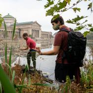
On a recent afternoon in the “Microbiomes Across Environments” course, taught by MBL faculty David Mark Welch and Elena Peredo, nine students were busily engaged in lab activities, conversing through face masks and across 6-foot distances. They had been out on the MBL’s research vessel, the Gemma, where they collected sea urchins, starfish, and horseshoe crabs.
Back in the lab, they used swabs to sample the microbiome from several organs of the animals, and talked about the research questions they might ask. Is the microbiome different between a male and female crab, and how? How do the mouth and anus microbiomes differ? How does the microbiome change after the animal has been in the lab for several days? “We are teaching them how microbial ecologists ask questions,” said Mark Welch.
“I was really excited about the field component of this course,” said Megan Garvey, a biology and visual arts major, “especially now when everything is online.”
McKibben agreed: “Interacting with the animals, doing the DNA swabs, was really unique. At another school, you might just get little tubes of samples. Here, you are a part of where the organisms actually came from.”
That day, the students worked on purifying and amplifying short pieces of DNA from the microbial samples. These pieces will be sequenced in the MBL’s Bay Paul Center, and the students will get back computer files containing tens of thousands of unprocessed DNA sequences. They will then use bioinformatics to analyze the data, so they can answer the comparative questions they’ve chosen.
“There’s been a real sea change in our understanding of what it means to be an organism,” Mark Welch said. “The microbiome is a critical part of an organism’s health and behavior, the way it develops, whether or not it can regenerate. All of the students, no matter what their major, will leave with that appreciation for the complexity of organisms.”
A lot to see
“Imaging for Biological Research” is a new course developed by MBL Director Nipam Patel and co-taught by Louis Kerr and Carsten Wolff of the MBL’s Central Microscopy Facility. Students learn the fundamentals of imaging, then quickly advance to using state-of-the-art microscopes to address biological questions.
Indeed, a few weeks into the course, students can sit comfortably at the controls of a $500,000 confocal microscope in Loeb Laboratory.
“We don’t usually get to use microscopes like this! Dissection microscopes are what you normally find in a lab,” said Jonathan Tang, a biology major, as he works intently to image a glowing fruit-fly embryo.
The four students are learning the careful work of identifying an embryo at a certain stage of development, mounting and orienting it correctly on a slide, and obtaining high-quality, beautiful images using the confocal. Each student is responsible for imaging two of the eight genes called Hox that are expressed along the head-tail axis of the embryo, Patel explained: “These genes have been known for a long time, but they are ideal for teaching students the fundamentals of developmental biology.”
Soon, they will move to imaging other organisms, including zebrafish, butterflies, crustaceans, and skates. And in the last three weeks, they will each have an independent project, guided by one of the faculty.
One student will work with Gayani Senevirathne, a postdoctoral course assistant from the University of Chicago, to image the Hox genes in zebrafish. Kerr will help a student image butterfly wing scales using electron microscopy. Wolff and a student will look at live cell movements in early crustacean embryos.
And Patel will help the fourth student image all eight Hox genes in a single embryo, which has not been published before.
“It will be really fun to see, and it will also make a great textbook figure,” said Patel, who plans to submit it to a journal. “More importantly, it teaches the students all the methods they need to get a figure like that.”
—Adapted from an article first posted by the Marine Biological Laboratory.