Government officials are required to keep records, so that newspapers or other officials can investigate when things go wrong—but cells have no such procedure. Scientists trying to understand cancer, for example, have to piece together clues and invent tools to understand what happened.
One such memo produced by cells that dissipates almost immediately is a molecule called nitric oxide, or NO. Scientists want to track this molecule’s movements, since it’s important in many disorders and diseases, particularly cancer. But to date, the only way to find out what NO was up to was to slash the whole cell open and count what comes out—losing all the information about where in the cell this molecule was operating.
A group of University of Chicago chemists, however, announced an innovative system to “spy” on NO as it goes about its business in live cells. Made out of DNA, the breakthrough probe lights up when it encounters NO within the cell, allowing researchers to better understand the molecule’s activities, develop drug targets, and check whether a drug is reaching its intended target in a cell.
“This is very exciting, both as a tool for understanding the fundamentals of how biology works and also as a way to improve drugs that are already in clinical use,” said Prof. Yamuna Krishnan, a leading biological chemist and co-author of a paper published March 9 in Nature Chemical Biology. “It’s the first way invented to track the molecule in living cells with sub-cellular accuracy.”
Scientists know that NOS3 - an enzyme that produces NO - spends most of its time in two major locations in the cell. One is at the cell membrane, the other is on an intestine-looking compartment called the Golgi apparatus that packages proteins. “The activity of NOS3 at these two locations is critical in disease, but it’s been very challenging to tell what NOS3 is doing and where,” said UChicago graduate student Maulik Jani, first author of the paper.
It was a perfect question for Krishnan’s lab, which specializes in building tiny sensors to travel inside cells and report back on what’s happening. To understand the workings of cells ranging from neurons to neutrophils, they build miniature cellular probes out of DNA—which the cell dissolves harmlessly after their job is done.
The team started with a package built out of DNA and attached several molecules: one that lights up in the presence of NO, plus a “mailing address” that sticks to either the cell membrane or the Golgi apparatus. In a nod to another gene technique, they called this sensor ‘NOckout.”
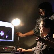
Once released in the cell, NOckout travels to its intended destination. The fluorescent alarm gets tripped whenever it encounters NO, and simply by looking for the glowing molecules under a microscope, scientists can see where in the cell NO is produced and how much of it there is.
The scientists are excited for its implications. The tool can help track what’s going on when NOS enzymes misbehave, as in cancer, heart disease and neurodegenerative diseases. “But you don’t always know whether the disease is due to too much or too little NO being produced,” Jani said.
“And if you already have a drug, this gives you a way to track whether your drug is actually working the way you think it’s working, which is often missing and is invaluable in pharmaceutical research,” Krishnan said.
In addition to the cell membrane and Golgi apparatus, Jani said, they already have mailing addresses that could be adapted to send probes to five other important cell organelles. And it could work for other hard-to-track molecules that perform important but mysterious cellular roles.
“NO is a very reactive molecule—it reacts with basically anything it encounters—so it’s difficult to catch in the act. There are lots of other molecules like NO that such an approach could be useful to track,” Jani said.
The other co-authors on the paper were graduate student Junyi Zhou and postdoctoral researcher Aneesh Veetil.
Citation: “A DNA-based fluorescent probe maps NOS3 activity with subcellular spatial resolution.” Jani et al., Nature Chemical Biology, March 9, 2020. https://doi.org/10.1038/s41589-020-0491-3
Funding: University of Pennsylvania Orphan Disease Center, Andrew Coppola Foundation, the University of Chicago Women’s Board, National Institute of Diabetes and Digestive and Kidney Diseases, the Chicago Biomedical Consortium, Searle Funds at The Chicago Community Trust, Brain Research Foundation, Mergel Funsky award.